Retinal detachment is a serious eye condition in animals where the retina detaches from the back of the eyeball, causing temporary, or permanent vision loss. The condition is usually caused by underlying health concerns of which some breeds are predisposed to.
What is a detached retina?
Separation of the retina from underlying tissues is known as retinal detachment. The retina is very thin tissue in the eye that is attached to a deeper underlying vascular layer and sits in front of a thick gel-like vitreous. Retinal detachment is a serious condition that can lead to permanent blindness if not treated promptly.
What is the retina?
The retina is a very thin tissue at the back of the eye that contains photoreceptor cells which can convert light into electrical signals. These signals then pass along nerve fibres, through the optic nerve and into the brain. Vision is the brain interpreting these signals.
Animals at risk of retinal detachment
Various breeds of cats and dogs are at particular risk of retinal detachment, due to their predisposition to conditions that allow retinal detachment to occur. These conditions include:
- Retinal dysplasia
- Collie eye anomaly
- Persistent hyperplastic primary vitreous
Dog breeds at particular risk of retinal detachment include:
- English Springer Spaniel
- Shih Tzu
- Samoyed
- Miniature Schnauzer
- Border Collies
- Labrador Retriever
- Bedlington Terrier
- American Cocker Spaniel
What causes retinal detachment?
Whilst retinal detachment can occur in any breed at any age, it mostly affects older dogs with underlying health conditions such as glaucoma and hypothyroidism. Other causes of retinal detachment can include:
- Congenital abnormalities
- Trauma
- Inflammation
- Infection
- High blood pressure
- Cancer
- Intraocular surgery
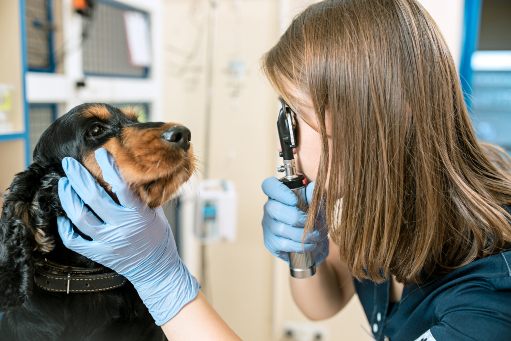
Diagnosis of retinal detachment
Distant direct ophthalmoscopy, direct ophthalmoscopy, slit-lamp examination and ultrasound may all be used in the diagnosis of retinal detachment. Tests may need to be performed and sent to a laboratory for analysis to determine associated diseases. Your vet may also need to perform various tests to determine the cause of retinal detachment in order to effectively treat the condition.
Treatment options for retinal detachment
Whilst laboratory tests are being analysed, a course of topical eye drops or tablets may be administered to encourage the retina to reattach itself. In less severe cases where the retina successfully reattaches itself, your vet will then treat any underlying causes.
If medication is not enough to reattach the retina, surgery may be required to regain vision. This may include surgical methods such as laser retinopexy and vitreo-retinal surgery at a specialist tertiary referral hospital.
Detached retina recovery
If surgery is required to reattach the retina, your vet will recommend that you restrict your pet’s activity in order to help the eye heal. Several follow-up appointments will also be required to monitor your pet’s health and ensure that there are no complications.
Knutsford Vets Surgery
By choosing Knutsford Vets Surgery, your pet will be in the safe hands of experienced Ophthalmologist, Dr Paul Adams. Dr Adams has years of experience treating a wide range of eye conditions, and is the perfect partner to look after your pet’s ocular health.
We welcome new patients, second options and referrals.
Our friendly team is on hand if you have any questions. Contact us or call us on 01565 337999.
It is important to note that if your dog has had Cherry Eye in one eye, they are at risk of developing it in the other. It is also important to keep your dog’s eyes moist. Check their tear production and use hydrating eye drops as necessary.