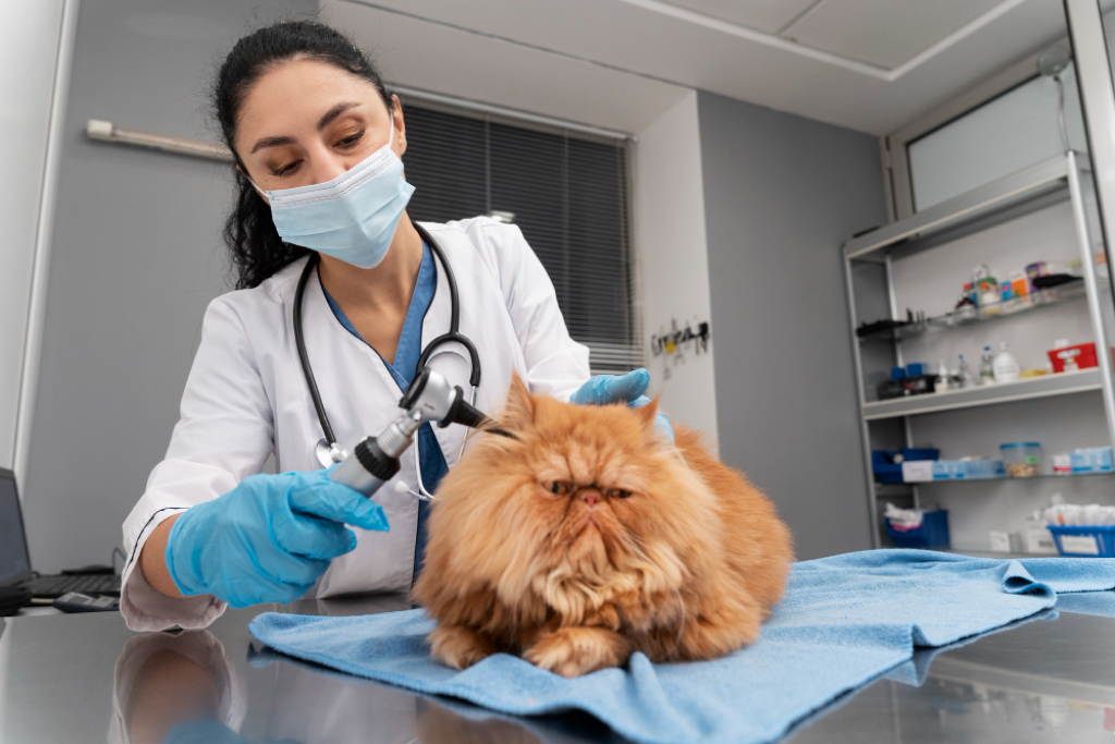
Understanding Diagnostic Tests
We understand that the diagnostic process can sometimes be long and stressful. But not knowing or understanding what’s happening could make the situation even worse. In this article, we go over the different types of diagnostic tests that your vet may decide to use when determining what’s wrong with your pet, as well as how these tests help your vet to plan a suitable course of treatment.
So, what are diagnostic tests for pets?
There are a wide range of diagnostic tests that vets use to determine what’s wrong with your pet, but cytology and histology are amongst the most common. Cytology studies the structure and function of your pet’s cells, whilst histology looks at their tissues, organs, and other bodily systems.
Read on to learn more about the diagnostic tests that vets use to work out what’s wrong with your pet, and to help determine the next steps, or a suitable treatment plan.
What is the Difference Between Cytology & Histology?
The main difference between cytology and histology is that cytology studies the characteristics of small groups of, or individual cells, whilst histology studies how cells relate to each other in thin slices of tissues or organs of the body. As such, cytology has a fairly narrow study area, whilst the study area of histology is much wider.
Cytology
What is Cytology?
Cytology (sometimes referred to as cytopathology), is the microscopic study of cell samples collected from your pet’s body; commonly via fine needle aspiration, or fine needle biopsy, both of which use a sterile, fine gauge needle. Skin scrapings, impression smears, and swabs are also commonly used to collect samples for cytology.
Vets use these samples to help diagnose growths or masses, but cytology can also be used to assess bodily fluids, internal organs, and abnormal fluids that may accumulate in the chest or abdomen.
Digital cytology can also be used to help speed up the analysis of cytology samples. Scopio is a top-of-the-range diagnostic tool we can use here at Knutsford Vets. Once we collect your pet’s cytology samples, we simply have to scan the slides into the Scopio system, and a remote pathologist will be able to provide a report within an hour.
Vets who still send slides to an external laboratory typically have to wait 7-10 days for results. With digital cytology, at Knutsford Veterinary Surgery we can treat your pets in real time, diagnosing quicker and starting treatment sooner.
When is Cytology Used?
Cytology is commonly used to diagnose the nature of ‘lumps and bumps’, but can also be used to assess:
- Internal organs, such as the liver, adrenal glands, and kidneys
- Bodily fluids, such as urine or joint fluid, as well as abnormal accumulations of fluids, particularly in the chest and abdomen
- Various surfaces of the body (internal and external), including the eyes, the mouth, and airways
Cytology samples collected from these areas can then go on to reveal if the problem is caused by inflammation, infection or an abnormal growth of tissue. However, if it is determined that abnormal tissue growth is the cause of the problem, cytology can usually determine which type of cells are involved and indicate if the growth is likely malignant or benign.
Advantages of Cytology:
Cytology has a number of advantages, making it a great diagnostic tool for vets. These include:
- Simple and quick, especially when digital cytology is used
- Minimally-invasive, relatively painless, and sampling can often be performed without sedation or anaesthesia
- Can provide a definitive diagnosis, or at least helps to determine the next steps
Limitations of Cytology:
Whilst cytology does have a lot of great diagnostic advantages, it falls somewhat short, in that it doesn’t always tell the whole story. Usually, this happens if there is an issue with sampling (if they are very small, or if the relevant diagnostic cells are not present in the sample). However, sometimes, the disease or tumour in question simply doesn’t present in the standard way, in which cases cytology may not be able to provide a clear diagnosis.
That being said, cytology will usually give vets an idea of what is going on, even if it doesn’t provide a definitive answer, and will indicate the next steps to follow. In unclear cases, the next step will usually be histology.
Histology
What is Histology?
Histology (sometimes referred to as histopathology) is the study of tissues, usually collected surgically. Samples are prepared by preserving, thinly slicing or sectioning, and staining with dyes before being examined under the microscope by a pathologist.
The accuracy of histology is typically fairly high, and the pathologist can usually offer diagnosis, as well as prognosis (i.e. what caused the disease). With this information, your vet can then begin to determine the best course of treatment for your pet.
When is Histology Used?
Histology is typically used when cytology cannot make a definitive diagnosis. It is used in the same cases as cytology, but it provides a more accurate level of detail in order to make a definitive diagnosis. What’s more, where tumours and cancers are concerned, histology will also provide tumour/cancer classification which will assist vets when determining treatment options.
Advantages of Histology:
The advantages of histology include:
- Fairly high diagnosis accuracy
- Can offer an opinion on the cause of the problem
- Large sections of tissue can be studied, providing the pathologist with more information to examine
- Samples usually require collection under general anaesthetic
- Pathologists may be able to offer an opinion as to whether removed tumours have a clean margin (i.e. if the entire tumour has been removed)
- It is an essential diagnostic tool, allowing vets and pathologist to examine the internal structure of of tissues
Limitations of Histology:
Whilst histology is typically the next step after inconclusive results from cytology,
it has own its limitations. A definitive diagnosis is still dependent on a viable sample, and sometimes it can be difficult to obtain a large enough sample. In this case, results are often inconclusive and a second biopsy is required.
How Does Cytology and Histology Help Vets Manage a Problem?
The results of cytology and histology are essential in helping your vet determine an appropriate course of action, and suitable treatment.
Cytology, results that indicate inflammation can signal your vet to begin looking for a specific cause, such as bacteria, parasites, or fungus, whereas results that indicate abnormal growths will often indicate the type of tumour your pet has, and will help to predict how it may behave. However, further tests, such as histology and chest x-rays may be required for more detailed analysis of the condition.
The results of histology help pathologists and vets to classify the tumour or cancer, and make an accurate diagnosis. Your vet will then use this information to plan a suitable course of action, treatment, and may help to predict the behaviour of the cancer.
How are Samples Collected?
Samples are collected in different ways for cytology and histology. Samples usually require collection under general anaesthetic. What’s more, cytology samples are usually non-invasive, relatively painless.
Cytology
Cytology samples can be collected in a number of different ways, depending on the area in question, and what your vet needs to learn. Common cytology sampling methods include:
Skin Scraping:
Skin scraping is used to rub cells from the surface of the skin, e.g. on a patch of flaky skin, a bald spot, or an ulcerated lump. A sterile scalpel is firmly dragged across the skin several times (not cutting into the skin) to scrape away the top layers of skin cells. This sample is then spread thinly onto a glass slide, stained with special dyes, and examined under a microscope, or digitally via tools such as Scopio.
Impression Smears:
Impressions smears are used when there is a draining or weeping wound. Vets collect the surface material (i.e. the liquid weeped) by pressing a glass slide against the area several times to build up enough material to examine under a microscope. Smears are useful for detecting the presence of inflammation, infectious organisms, and abnormal tissue cells.
Swabs:
A cotton-tipped swab (a bit like a Q-Tip) is used to collect cells from internal surfaces, such as inside the mouth, eye, ear, nostril, prepuce, and vagina. The swab is then rolled against a glass slide, and examined under a microscope. Results often reveal inflammatory cells, infectious organisms, and tissue cells from the surface that can be examined for signs of abnormality.
Fine Needle Aspiration/Fine Needle Biopsy:
This technique involves using a sterile, fine gauge needle, which is attached to an empty syringe. The needle is inserted into the area in question and suction is applied to withdraw cells from inside that area. This method is often used on solid tissues, such as organs or tumours, and fluid-filled tissues, such as joints or cysts.
What Happens Next?
After samples have been collected, a pathologist will examine them and offer their opinion, diagnosis, and prognosis (where possible). If tests come back inconclusive, the information gained will help vets to plan a suitable course of action, including further testing and treatment options.
Introducing Scopio & How It Can Help to Detect Cancerous Cells
Scopio is an all-in-one digital cytology tool that offers results within an hour. At Knutsford Vets, we use Scopio to bring you much needed answers much quicker than usual, and to get the information that we need in order to plan a suitable course of treatment.
What is Scopio?
Scopio is a digital cytology system that allows vets to send high-resolution images of your pet’s sample to a remote pathologist for examination. By using Scopio, we get results within an hour, meaning that we can begin planning your pet’s treatment or next steps the very same day. What’s more, Scopio is available 24/7.
How Does it Work?
We scan your pet’s sample slides into the Scopio system, which are then digitally sent to a certified remote pathologist for examination. Results are returned within an hour to allow for same-day continuation of care – this means that we can get a head start on planning and treating your pet before leaving the clinic.
What Does This Mean For My Pet?
The key benefit of using Scopio is that it provides vets with critical information about your pet’s condition sooner than the usual testing methods which can take days. Scopio lets us know more about your pet’s condition, sooner, so that we can begin caring for your pet much quicker.
Scopio offers:
Results within an hour
24/7 access to board certified pathologists from around the world
Same-day care and treatment
Pet Testing & Diagnosis at Knutsford Vets
At Knutsford Vets, we have a range of in-house diagnostic tools that help us to provide answers quickly and accurately so that we can get a head start on treating your pet. If your pet has suddenly started acting unusually, or you have noticed some concerning symptoms, get in touch with us, or book an appointment today.
Our vets will examine your pet, take any necessary samples, and work to provide you with answers and a course of action as soon as possible.
